Vote Here ~~~~~~~~~~~~~~~~>>>>>>>>>>
CASE HISTORY:
Sasha is a Domestic Medium Haired feline, 12.5# and 6.5 yrs old. She was referred to us by Dr. Bob Mills of North Arkansas Veterinary Clinic. Sasha had a soft tissue mass/cyst involving left caudal 2 mammary glands.
There were previous attempts at drainage, and the biopsy had yielded suggestion of carcinoma. Dr. Dew discussed a resection of abnormal tissue and submission for histopathology under assumption the diagnosis will be a mammary carcinoma.
There were previous attempts at drainage, and the biopsy had yielded suggestion of carcinoma. Dr. Dew discussed a resection of abnormal tissue and submission for histopathology under assumption the diagnosis will be a mammary carcinoma.
TREATMENT:
Dr. Dew used a ventral approach and performed a mass excision on Sasha's left two mammary glands. The mass size was 10 x 7 x 3 centimeters, and extended to her midline. There was no deep or lateral extension noted. Her prognosis is good for soft tissue healing. The use of the e-collar, confinement and restricted activity will be required to decrease the likelihood of complications. The long term prognosis will be discussed after the biopsy results are returned.
CASE HISTORY: Cinnamon
 Cinnamon is a Boxer Mix. She is 3-1/2 ys old and weighs 53.8#. She was referred to us by Dr. Hartford Hamilton with Pleasant Valley Veterinary Clinic last year for a Left TTA in July. Upon physical exam, Cinnamon is doing well on her left TTA, and is now acutely lame on the right. Findings reveal that she is in need of a Right TTA.
Dr. Dew used a medial approach for Cinnamon's TTA (Tibial Tuberosity Advancement) surgery, and observes that her meniscus is also torn and is at Grade II. Harware used in her knee consists of a 9x19mm basket, 5 hole plate/fork, and 4 titanium screws obtained from Kyon in Zurich, Switzerland. A bone graft is also placed beneath the basket to promote healing.
Tucker
Tucker is a little Yorkie who weighs 3# and is 4yrs and 2 mos old. Dr. David Oates from Russellville Animal Clinic has sent him to us. According to his owner, Tucker has always been quite a sedate puppy. Recently bouts of forelimb paddling had been noted which have resolved with lactulose. Bile acids completed with Dr. Oates where normal preprandial and over 70 postprandial. BUN was not available prior to today. Dr. Dew discussed a high probability of a PSS, also microvascular dysplasia. Tucker's owner wished to proceed with abdominal exploratory and biopsy of vascular occlusion.
TREATMENT:
Dr. Dew approaches Tucker's abdominal exploratory surgery ventrally. He sees no intra or extrahepatic shunt, and the portal vein is of normal size. He then performs 2 hepatic biopsies, one on the right quadrant and one on the left. Dr. Dew observes definite hepatic dysfunction, and he suspects microvascular dysplasia. The prognosis for uncomplicated soft tissue healing is good. The long term prognosis will be discussed after the biopsy results are returned.Jack
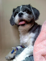
Jack is a 2 yr 4 mo, 14# Shih Tzu. Dr. Lynn Rodgers of Huntsville Veterinary Clinic sent him to us for eval and possible surgery when he had an acute onset of rear limb weakness on the Thursday prior. On Friday, he lost his ability to ambulate. He was treated with steroids but there was no improvement. Upon exam with Dr. Dew, he had Grade 4.5 rear limb neuropathy; there is deep pain present but significantly decreases in left, and is not present on right. Dr. Dew discussed severity of signs, and the prognosis with immediate surgery, and the possibility for neurologic improvement for up to 6 months. His owners wished to proceed with CSF, myelogram and hemilaminectomy if indicated.
TREATMENT:Cerebrospinal centesis: There was 2ml clear CSF collected from cisterna magna.
Myelogram: Injecting 3ml iohexol, column thinning was visible on x-rays from L2-L5, and there was a slight deviation of his spinal cord to left at L3-4.
Hemilaminectomy: Dr. Dew used a dorsolateral approach to the Lumbar 2-3 and 3-4 discs. He removed a large amount of clot and acute disc material, and observed some cord discoloration under the dura. Jack was doing very well the day after the hemilaminectomy procedure and was using both rear legs with the help of Tail-walking.
Butkus
Butkus is a 110#, 1 yr 2 mo old Rottweiller who comes to us from Dr. Bob Hale at Briarwood Animal Hospital. He had his Right THR (Total Hip Replacement) done by us back in September 2009, and he is doing great on the right THR. There was significant improvement in his muscle mass. He returned to us for a Left THR.
TREATMENT:
Dr. Dew used a craniolateral approach for Butkus's surgery. He prepared Butkus's acetabulum and femoral canal in the standard fashion. The acetabular cup (part of the implant for THR) perforations were packed with a bone graft. The implants included a 29mm acetabular cup, medium femoral stem, 28mm stem screw, and a short neck obtained through Kyon. Butkus has an excellent prognosis for an uncomplicated recovery and pain free right hip function. Over the long term, the complication rate for cementless total hip replacement is 4-7%. Many of the early complications (hip luxation and femoral fracture) can be greatly decreased with exercise restriction and nursing care.
Joey
Joey is a Pembroke Welsh Corgie weighing 36# and is 6yr and 3mo old. Dr. Todd Kollasch at Greene Animal Hospital sent him to us because of a history of urethral blockage due to calculi in September. Dr. Kollasch completed a scrotal urethrostomy and cystotomy with stone analysis. Due to an issue with pancreatitis "Joey" was not placed on S/D diet post-operatively. Over the weekend prior to meeting with Dr. Dew, stranguria developed. Dr. Kollasch was able to pass a small bore catheter, but he wishes to have Joey's urethra and bladder assessed. His owner utilized the Azzore transport service from the Memphis area.
TREATMENT:Dr. Dew performed a urethragram. The survey radiographs demonstrate multiple cystic calculi. There was no evidence of stricture, as the urethragram was completed with a foley catheter. After a quick call to Joey's owner, Dr.Dew then performed a cystotomy, calculi removal and analysis, an abdominal exploratory and bile acid assessment. There was no evidence of a shunt vessel during the abdominal exploratory with ventral approach. There was an omental mass sizing 3x2cm (Dr. Dew suspects this may be an accessory spleen), the bladder was slightly thickened, and multiple spiculated calculi were removed. The calculi obtained were sent to the state lab for analysis.
Hobo
CASE HISTORY:
Hobo is a 77.5#, 6yr old Doberman. He was referred to us by Terry Barnett at Millington Animal Hospital. Trauma had resulted in a fracture of one of his phalanges (a digit) on his paw. His owner wished to have him transported from Memphis to Russellville for surgery.
TREATMENT:
Dr. Dew used a dorsolateral approach for Hobo's surgery. He was able to provide good reduction and hardware placement. Hardware used were 0.045 and 0.035 thread pins. Prognosis is good for normal osseous healing. Exercise restriction and bandage care instructions must be followed to maximize the prognosis for uncomplicated healing.
Sammi
Sammi is a 50# 2yr old Boxer sent by Carl Fulton of Faulkner County Veterinary Clinic. She was hit by a 4-wheeler, which resulted in the luxation of her Right Tarsal/Metatarsal joint. She has been treated with splinting for the past 7 weeks, but lameness and instability persist. Dr. Dew suggested partial Tarsal Arthrodesis utilizing ESF (External Stabilization Fixator) and bone graft. Sammi's owner decided to proceed arthrodesis today.
TREATMENT:Dr. Dew used a medial to proximal tibia approach on this surgery. He was able to give good alignment and good cancellous graft placement. He used 4 small enhanced threads and 2 smooth pins along with acrylic sidebars to create the ESF.There is a good prognosis for Sammi's return to weight bearing function in 16-26 weeks.
Brandy
Brandy is a Pomeranian. She is 5-1/2 years old and weighs 11.9#. She comes from Dr. David Oates at the Russellville Animal Clinic. She had an acute onset of rear limb paresis after falling from bed the night before. Dr. Dew observed a Grade IV-V rear limb paresis, with panniculus evident at T10. There was deep pain present in her Left rear but her response was blunted. Her owner wished to proceed with Myelogram and Hemilaminectomy surgery if indicated.
TREATMENT:Cerebrospinal centesis: As this was obvious trauma with known cause, CSF fluid was not obtained.
Myelogram: Injecting 4ml iohexol, column thinning was visible on x-rays from T13-L1, and there was a slight deviation of his spinal cord to left at T12 - L1.Surgery will be beneficial, but due to cord swelling prognosis is guarded until improvement is noted.
Hemilaminectomy: Dr. Dew used a dorsolateral approach at T13. He observed the cord to be swollen, and there was an old hemorrhage at the site. Some acute disc material was removed. With the severity of Brandy's clinical signs, a transient worsening of neurologic function can be noted after a myelogram and surgery. This condition will usually resolve to the pre-operative state in 3-10 days. The nervous system is very slow to heal. Improvement can be expected for up to 5 months. Nursing care, physical therapy and exercise restriction all play critical roles in avoiding complications and aiding recovery.


No comments:
Post a Comment