Thursday, November 19, 2009
A busy day ahead for the Dew Crew! Five post-surgery rechecks, three procedures and four surgeries.
CASE HISTORY:
 Brownie came to us at the beginning of the month for rear limb paresis (paralysis). He had been treated over the weekend for sudden onset of pain and then paralysis over the weekend at the Fort Smith Animal Emergency Center with no improvement. He came to Azzore Veterinary Specialists on Wednesday unable to walk or support himself at all on his hind legs, but deep pain was still present. Prognosis for Brownie was fair post surgery.
TREATMENT:
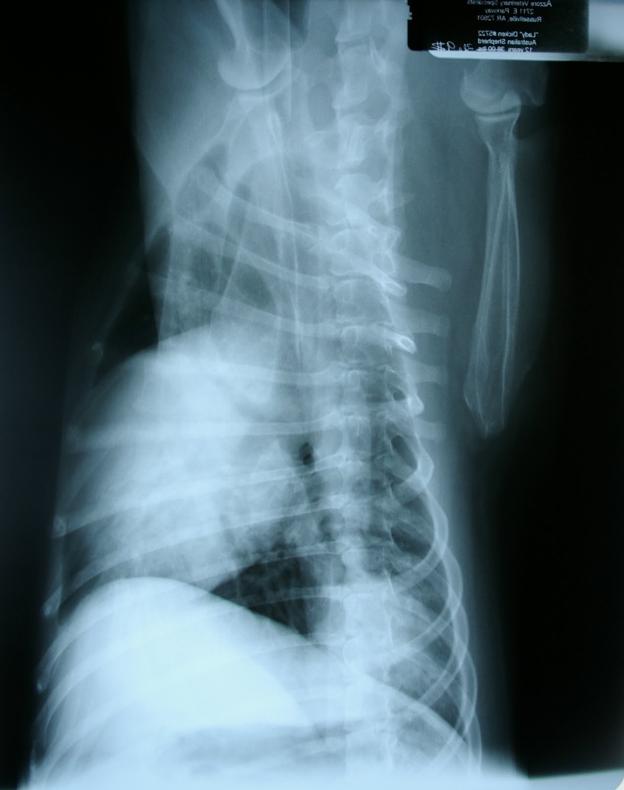
Myelogram: X-rays with contrast medium (iohexol) showed column thinning from L3-L5 and a slight deviation of the cord to the left at L4-5. While Dr. Dew determined that a hemilaminectomy would be beneficial, due to cord swelling, Brownie's prognosis was downgraded to guarded.
Hemilaminectomy: A large amount of disc material was removed at L4-5, and durotomy (incision to the outer covering of the spinal cord) performed to relieve pressure.
Hemilaminectomy: A large amount of disc material was removed at L4-5, and durotomy (incision to the outer covering of the spinal cord) performed to relieve pressure.
RESULT:
Two weeks after Brownie's surgery, he is standing without assistance on his hind legs! Mom says he is getting around some at home. Although we observed some knuckling of his left rear paw and he is still non-weight bearing on his right leg, he is far ahead of our expectations based on his pre-surgical condition. CASE HISTORY:
We first met Abraham at the beginning of September three days after he had been hit by a car. Abraham suffered a spinal fracture (L6), couldn't walk, but deep pain was still present in both rear limbs. His prognosis for neurological recovery was only fair.
TREATMENT:
Abraham's spine was stabilized using a technique that Dr. Dew helped to develop, using pins and an external fixator with acrylic bars.
RESULT:
2 1/2 months post surgery, Dr. Dew removed Abraham's external fixator on Thursday. He is walking well with no signs of discomfort -- and really, really happy to have that thing off of his back!
CASE HISTORY:
Lady was back for her one week recheck. Lady came to our surgery center for a left caudal lung lobectomy due to a mass.
The mass was confined to middle of the left caudal lobe. Tissue was sent to the lab for analysis. Pending lab results, Lady's prognosis was good.
RESULT:
One week post surgery, Lady's incision is healed and she is breathing normally. Lab results show pulmonary adenocarcinoma with moderate mitotic activity.

CASE HISTORY:
Remember Spitfire? Spitfire the Weimaraner came to Azzore for progressive right forelimb lameness and valgus deviation (medical jargon meaning that instead of her leg being straight up and down, it angled out to the side). Dr. Dew's diagnosis: infraspinatus contracture of the tendon in Spitfire's shoulder.
TREATMENT:
Infraspinatus tendon release via tenotomy (surgical procedure that cuts the tendon of a contracted muscle to allow lengthening).
RESULT:
Two weeks post surgery, Spitfire is walking almost normally. We have video footage of before and after which we will try to get up on YouTube this week so that you can see the amazing difference in Spitfire's gait!

Quincy's veterinarian referred him to Azzore after he experienced acute onset of right rear lameness. Dr. Brown identified a cranial cruciate ligament injury.
TREATMENT:
Dr. Dew corrected Quincy's injury with tibial tuberosity advancement surgery. In this procedure, the knee is biomechanically altered by moving the tibial tuberosity and attached straight patellar ligament forward. The piece of bone that is moved is stabilized by a permanent titanium implant system from Kyon.
RESULT:
Two weeks after surgery, Quincy's incision is healed and he is weight-bearing on the operated limb. Now comes the tough part -- his family has to keep him exercise restricted for another 10 weeks!

CASE HISTORY:
Voodoo first came to Azzore back in June for hyper extension of the left hock secondary to calcaneal tendon disruption (in plain English, a traumatic injury to his Achilles tendon).
TREATMENT:
This is a difficult injury to repair, and Voodoo had only a 70% prognosis for uncomplicated healing and normal use of his hock (ankle) in the future. The "fix" would include surgery to suture the tendon and to place an external fixator to unload the repair (reduce weight on the tendon) while it healed. But, the external fixator would have to remain in place for as long as possible -- in this case, 5 months!
RESULT:
Voodoo's external fixator was removed, and the hock and tendon were stable when manipulated during the exam. Three more weeks of leash restriction and a round of antibiotics, and Voodoo will be running with the wolves... or maybe just with the dogs...

Meet Roger. Roger experienced acute rear limb weakness on Monday starting on the left side. After medical management with his veterinarian, Roger has been referred to Dr. Dew for further treatment. He is weak in both hind limbs, but has good pain response on both sides and in both limbs. Roger was admitted Thursday for surgery on Friday.
TREATMENT:
Cerebrospinal centesis: Spinal fluid was collected from the cisterna magna prior to injecting the contrast medium.
Myelogram: a series of x-rays showed column thinning from T11-L1 and a slight deviation of the cord to the right at T11-12. Because of cord swelling, his prognosis is guarded. Roger's parents want to go ahead with surgery.
Hemilaminectomy: T11-12, T12-L1, left, disc material was removed.
RESULTS:
Time will tell. Often in these cases, the symptoms actually get worse right after the myelogram and hemilaminectomy. In most cases, there is a gradual return to the pre-surgical condition over the 3-10 days immediately postop. But, Roger still won't be out of the woods then. It can take 5 months of nursing care, physical therapy and exercise restriction to see just how much mobility is returned after neurological surgery. We'll let you know how Roger is doing after his 2 week recheck appointment!

Chloe was transported to our surgery center from Memphis, Tennessee. She suffered a traumatic rupture of her right cranial cruciate ligament when she was hit by a car. Chloe was admitted Tuesday night for surgery on Thursday.
TREATMENT:
Chloe’s injury was corrected with tibial tuberosity advancement surgery.
RESULT:
Post surgery x-rays show excellent hardware positioning. Chloe’s family will need to keep her exercise restricted during the 12-week recovery period to ensure complete healing.

Junior came to see Dr. Dew for a large open hygroma on his left elbow. What’s a hygroma? I’m glad you asked! In layman's terms, a hygroma is a swelling that develops – especially on elbows – when they repeatedly get smacked on a hard surface, or even from always lying on hard surfaces. This is particularly common in large breed dogs like Junior (Mastiff). The area becomes inflamed, and a dense-walled, fluid-filled cavity develops. Junior’s case was a particularly nasty one which Dr. Dew felt would require staged procedures to correct.
TREATMENT:
Junior’s wound was closed and the limb bandaged.
RESULT:
Time will tell. This will likely require multiple surgeries to correct.
P.S. I would have posted a picture of Junior's elbow, but thought I'd better not in consideration for those who read our blog and might be a bit on the squeamish side!

Maggie has suffered from tracheal collapse for years. Over the weekend, she suffered acute decompensation which required emergency treatment and medical therapy. X-rays taken by her veterinarian showed collapse of intra and extrathoracic trachea. Maggie was truly in a fight for her life and a race against the clock. Dr. Dew placed a temporary tracheostomy tube at McGehee Animal Clinic before transporting Maggie to our Russellville surgery center. A call was placed to special order an intratracheal stent in order to save Maggie’s life.
TREATMENT:
Maggie experienced an immediate improvement after placement of the tracheostomy tube. The intratracheal stent arrived Thursday morning. The Dew crew took a series of x-rays of Maggie to determine proper placement for the stent.
RESULT:
One day post surgery, Maggie’s breathing was greatly improved with only minor coughing (which is typical for a dog with a tube permanently placed in her windpipe!). We look forward to seeing Maggie at her post-op appointment in a couple of weeks.

Duke came to us Thursday after two months of left rear lameness. A cruciate ligament injury was suspected. However, x-rays of the stifles (knees) and hips revealed nothing. Dr. Dew detected a hock abnormality during the examination, and stress x-rays of Duke’s left hock (ankle) confirmed an Achilles tendon injury (see Voodo above!). It really shouldn't have that much "bend" to it.
TREATMENT:
Duke went to surgery Friday. His hock was stabilized with an external fixator comprised of pins and acrylic bars.
RESULT:
Unknown. Now comes the tough part! Duke’s family will need to keep him exercise restricted to on-leash activity only for six months to allow the tendon time to heal.


I bet Abraham was ready to get that thing off his back! Y-OUCH!
ReplyDelete